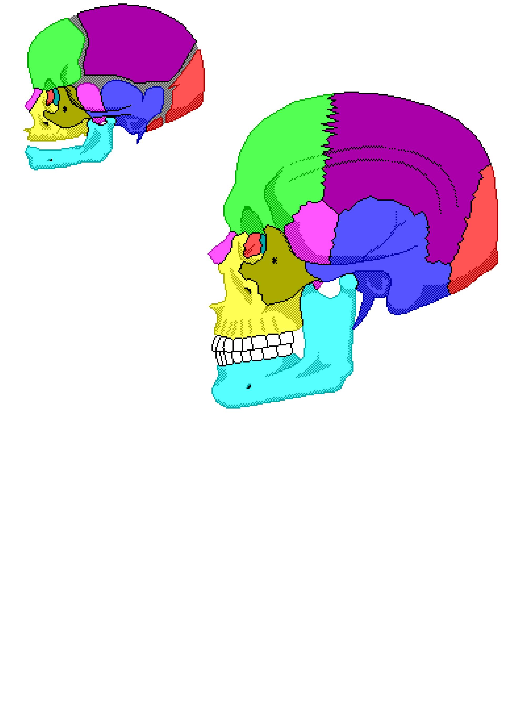
Human Skulls (Baby & Adult, Side View)
See Also: Spine Vertebra and Disk | Skulls of Baby and Adult (Above) | Did you know?

Viewing the Images

Anterior Fontanelle

The anterior fontanelle is one of the two areas of an infant's head where the skull bones have not completely covered the brain. It is typically known as one of the "soft spots." The anterior fontanelle is located towards the front of the head. The anterior fontanelle can normally be felt until 9-16 months of age.

Parietal Bone

One "parietal bone" is located on each side of the skull just behind the frontal bone. It is shaped like a curved plate and has four borders. Together, the parietal bones form the bulging sides and roof of the cranium. They are fused in the middle along the "sagittal suture," and they meet the frontal bone along the "coronal suture." Where the two sutures meet is called the "bregma." There is a gap through the parietal bone that serves as a passageway for blood vessels and nerves, called the "parietal foramen."

Coronoid Process

The coronoid process is one of the two processes the rami (lower jaw) is divided into. This thin triangular eminence serves as attachments for muscles used in chewing and can vary insize.

Ethmoid Bone

The "ethmoid bone" is located in front of the sphenoid bone. It consists of two masses, one on each side of the nasal cavity, which are joined horizontally by thin "cribriform plates." These plates form part of the roof of the nasal cavity, and nerves (ethmoidal cells) associated with the sense of smell pass through tiny openings in them. Portions of the ethmoid bone also form sections of the cranial floor, eye sockets, and nasal cavity walls. A "perpendicular plate" projects downward in the middle from the cribriform plates to form the bulk of the nasal septum. Delicate scroll-shaped plates called "superior" and "middle nasal conchae" project inward from the sides of the ethmoid bone toward the perpendicular plate. These bones, which are called the "turbinate bones," support mucous membranes that line the nasal cavity.

External Acoustic Meatus

The external acoustic meatus is a gap located at the lower edge of the temporal bones which leads inward to parts of the ear.

Fossa of Lacrimal Sac

The fossa of lacrimal sac is the vacant region between the frontal process of the maxilla and the lacrimal bone. It helps to lodge the lacrimal sac.

Frontal Bone

The "frontal bone" forms the front portion of the skull above the eyes and includes the forehead, the roof of the nasal cavity, and the roofs of the orbits (bony sockets) of the eyes. On the upper margin of each orbit, the frontal bone is marked by a "supraforamen" or ("suproorbital notch" in some skulls), through which blood vessels and nerves pass to the tissues of the forehead. Within the frontal bone are two "frontal sinuses," one above each eye near the midline.

Glabella

The glabella is the flattened triangular elevation that joins the superciliary arches. It is the flattened portion of the frontal bone.

Groove for Middle Temporal Artery

The middle temporal artery; also known as the deep temporal artery, is one of two branches of the maxillary artery that supply blood to the temporalis and anastomose. A branch of middle temporal artery can be pulpated just in front of the earlobe.

Head of Condylar Process

The head of the condylar process is the upper portion of the condylar process. It occupies an oval surface with a fibrocartliage covering for articulation at the mandibular fossa with the temporal bone.

Temporal Bones

A temporal bone on each side of the skull joins the parietal bone along the "squamosal suture." The temporal bones form parts of the sides and base of the cranium. The superior temporal line is the topmost curved line on the parietal bone and lies above the inferior temporal line. This is where the temporal fascia attach. The inferior temporal line also lies on the parietal bone, and is the site of attachment of the temporal muscle. Located near the lower edge is a gap, the "external auditory meatus," which leads inward to parts of the ear. The temporal bones house the internal structures of the ear and have depressions, the "mandibular fossae," that join the processes of the mandible. Below each external auditory meatus, there are two projections: a rounded "mastoid process" and a long, pointed "styloid process." The mastoid process provides an attachment for certain muscles of the neck, while the styloid process serves as an anchorage for muscles associated with the tongue and pharynx. The mastoid foramen is a prominent opening in the temporal bone which is located behind (or posterior) to the mastoid process. Generally, it provides the opening through which a vein and artery pass. A "zygomatic process" projects from the front of the temporal bone in the region of the external auditory meatus. It joins the zygomatic bone and helps form the prominence of the cheek.

Infraorbital Foramen

The infraorbital foramen is above the fossa at the end of the infraorbital canal. It creates a perforation in the maxilla near the inferior margin and transfers the infraorbital vessels and nerve.

Lacrimal Bones

A lacrimal bone is a thin, scalelike structure located in the middle wall of each eye socket between the ethmoid bone and the maxilla. A groove in the front leads from the orbit to the nasal cavity and provides a pathway for a tube that carries tears from the eye to the nasal cavity.

Occipital Bone

The "occipital bone" joins the parietal bones along the "lambdoidal suture." It forms the back of the skull and the base of the cranium. There is a large opening on its lower surface called the "foramen magnum," through which nerve fibers from the brain pass and enter the vertebral canal to become part of the spinal cord. Rounded processes called "occipital condyles," which are located on each side of the foramen magnum, unite with the first vertebra of the spinal column. The junction of the sagitall and lambdoid sutures is called the "lambda."

Posterior Fontanelle

The posterior fontanelle is one of two areas of an infants head where the skull bones have not completely covered the brain. It is typically known as one of the "soft spots." The posterior fontanelle is located at the upper back part of the head. The posterior fontanelle is no longer obvious when the infant is four months old.

Sphenoid Bone

The "sphenoid bone" is wedged between several other bones in the front of the cranium. It consists of a central part and two wing-like structures that extend sideways toward each side of the skull. This bone helps form the base of the cranium, the sides of the skull, and the floors and sides of the orbits (eye sockets). Along the middle, within the cranial cavity, a portion of the sphenoid bone rises up and forms a saddle-shaped mass called "sella turcica" (Turk's saddle). The depression of this saddle is occupied by the pituitary gland, which hangs from the base of the brain by a stalk. The sphenoid bone also contains two "sphenoidal sinuses," which lie side by side and are separated by a bony septum that projects downward into the nasal cavity.

Fontanels

At birth, the skull is incompletely developed, and the cranial bones are separated by fibrous membranes. These membranous areas are called "fontanels" or, more commonly, "soft spots." They permit some movement between the bones, so that the developing skull is partially compressible and can slightly change shape. This action enables an infant's skull to pass more easily through the birth canal. Eventually the fontanels close as the cranial bones grow together. The posterior fontanel usually closes about two months after birth; the sphenoid fontanel closes at about three months, the mastoid fontanel closes near the end of the first year, but the anterior one may not close until the middle or end of the second year.

The Tooth

A tooth is a hard structure, set in the upper or lower jaw, that is used for chewing food. Teeth also give shape to the face and aid in the process of speaking clearly. The enamel that covers the crown (the part above the gum) in each tooth can be broken down by acids produced by the mouth for digestive purposes. This process is called "decay". To prevent decay, good oral hygiene, consisting of daily brushing and flossing, is necessary. The hardest substance in the human body is one of the four kinds of tissue which make up the tooth. It is enamel and covers the crown (area above the gum line) of the tooth. A bony material called "cementum" covers the root, which fits into the jaw socket and is joined to it with membranes. "Dentin" is found under the enamel and the cementum, and this material forms the largest part of the tooth. At the heart of each tooth is living "pulp," which contains nerves, connective tissues, blood vessels and lymphatics. When a person gets a toothache, the pulp is what hurts.

Zgyomatic Bones

The zygomatic bones (sometimes called malar bones) are responsible for the prominences of the cheeks below and to the sides of the eyes. These bones also help form the lateral walls and floors of the orbits (eye). Each bone has a "temporal process," which extends down the back to join the zygomatic process of a temporal bone. Together, these processes form a "zygomatic arch." The smooth prominence between the eyebrows is called the "glabella."

Zygomaticofacial Foramen

The zygomaticotemporal foramen near the center of the temporal surface perforates the zygomatic bone. It serves as the transmitor of the zygomaticotemporal nerve.